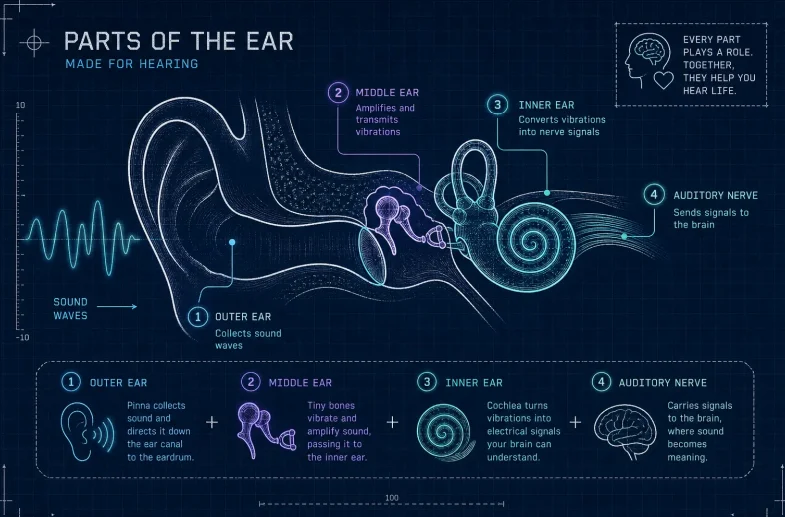
Key parts of the ear and how each affects hearing
The parts of the ear divide into three distinct regions, the outer ear, the middle ear and the inner ear. Each one handles a completely distinct job in turning sound waves into something your brain can understand. The outer ear captures and funnels sound, the middle ear amplifies it mechanically, and the inner ear converts those vibrations into electrical nerve signals. Beyond hearing, the inner ear also governs your sense of balance, which means a single compact structure does two remarkably important things at once. VR Speech and Hearing Clinic's certified audiologists provide free home hearing tests. This article covers the anatomy and function of each region, the conditions that affect them, and what all of this means if you are already wearing or considering a hearing aid.
Key takeaways
Outer ear functions:The pinna, ear canal and eardrum collect, channel and transmit sound waves to the middle ear.
Middle ear mechanics:Three tiny bones (malleus, incus and stapes) amplify sound pressure by roughly 22 times before passing it to the inner ear.
Inner ear roles:The cochlea converts vibrations into nerve signals while the semicircular canals and vestibule govern balance.
Condition mapping:Earwax blockage and swimmer's ear affect the outer ear; otitis media and otosclerosis affect the middle ear; sensorineural loss and Menière's disease affect the inner ear.
Hearing aid fit:The style of hearing aid you wear (BTE, RIC or ITE) is directly tied to which part of the ear it uses.
Red-flag symptoms:Sudden hearing loss, severe vertigo or pain behind the ear all need prompt medical attention.
How the parts of the ear collect and direct sound

The pinna's clever shape and why it matters
Think of the pinna, the visible, folded structure on the side of your head, as a cleverly shaped satellite dish for sound. Its irregular ridges and curves are not random decoration; they subtly shift certain sound frequencies before they even enter the canal, helping your brain work out whether a sound is coming from above, below or behind you. This directional function is easy to take for granted, which is why conditions such as congenital malformation or trauma that alter the shape or position of the outer ear can affect how well you localise sound in space.
The ear canal and what earwax really does
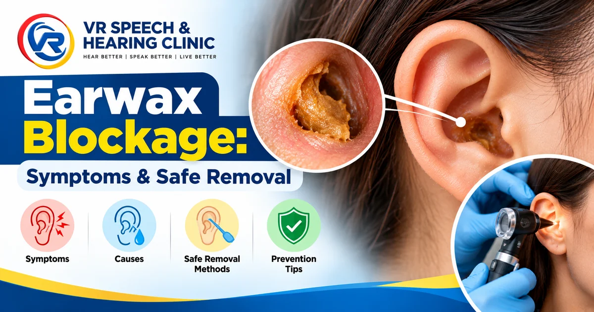
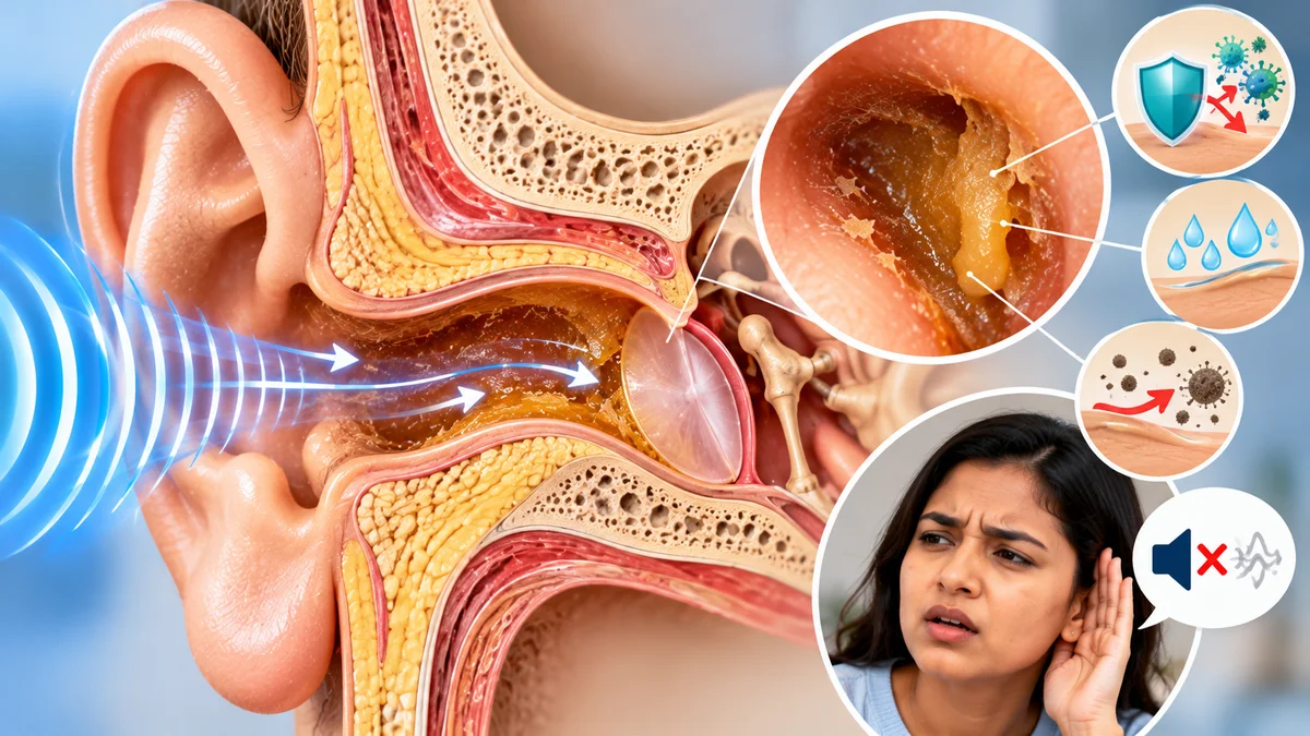
The external auditory canal is a slightly curved passage that channels sound toward the eardrum and mildly amplifies it along the way. Cerumen, better known as earwax, lines this canal and serves as a protective and lubricating barrier rather than a sign of poor hygiene. Too much earwax can muffle sound significantly, and this is one of the most common and most easily fixed causes of hearing difficulties. If everything suddenly sounds as though you are listening through a pillow but there is no pain, earwax build-up is often the first place to check.
The eardrum: where outer meets middle
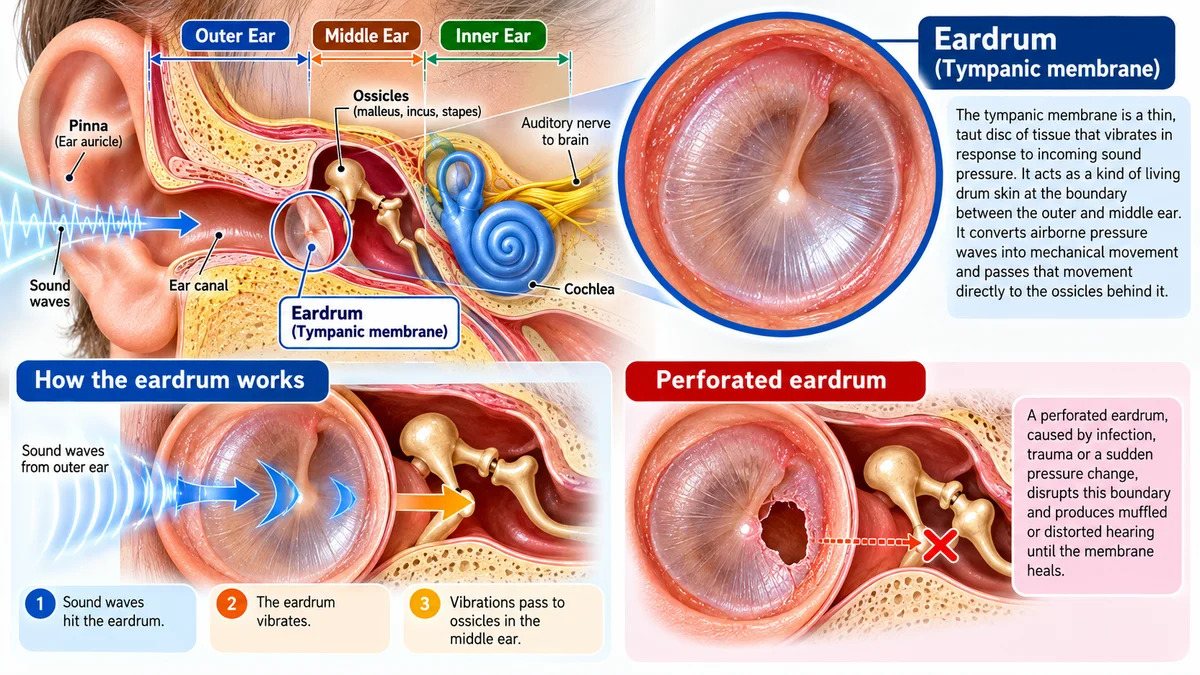
The tympanic membrane is a thin, taut disc of tissue that vibrates in response to incoming sound pressure, acting as a kind of living drum skin at the boundary between the outer and middle ear. It converts airborne pressure waves into mechanical movement and passes that movement directly to the ossicles behind it. A perforated eardrum, caused by infection, trauma or a sudden pressure change, disrupts this boundary and produces muffled or distorted hearing until the membrane heals.
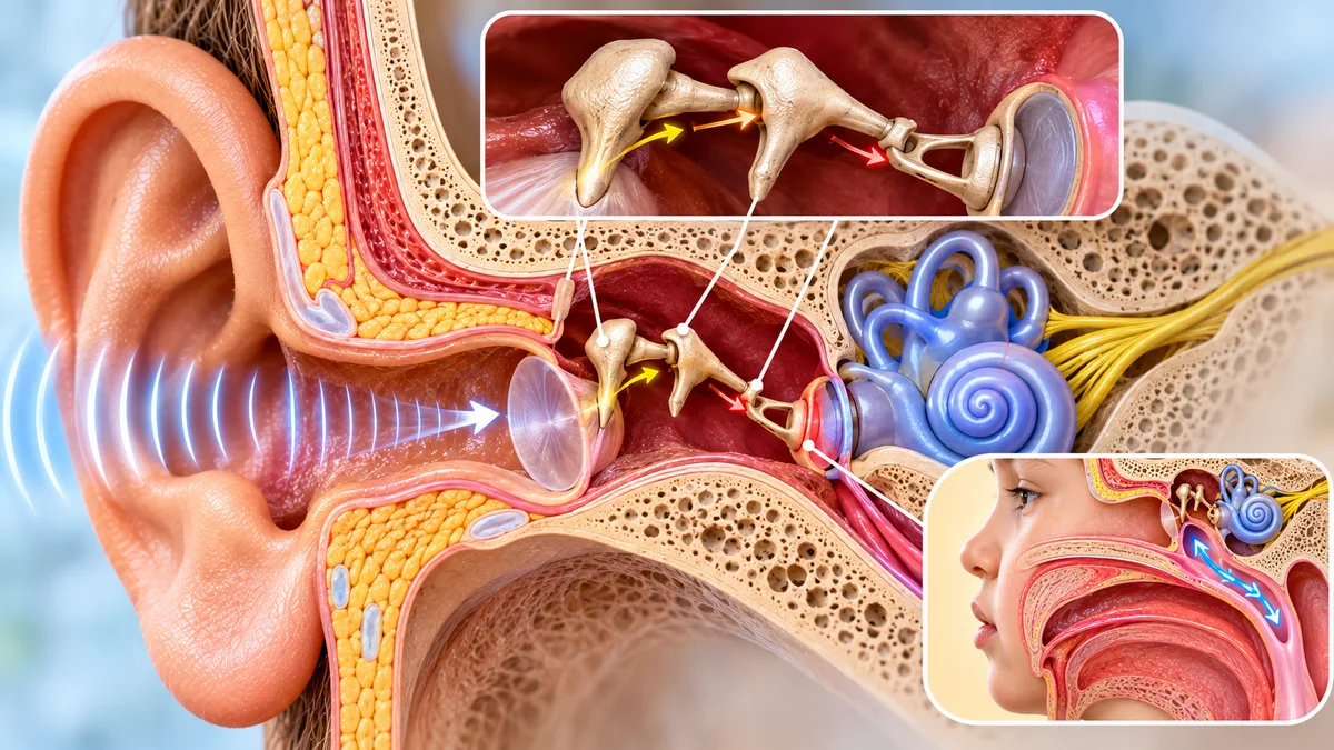
Parts of the ear: what the middle ear does
The ossicles: three tiny bones with a surprisingly large job
Behind the eardrum sits an air-filled space containing the three smallest bones in the human body: the malleus (hammer), the incus (anvil) and the stapes (stirrup), named for the objects they resemble. The chain reaction is elegant: the eardrum's vibration rocks the malleus, which nudges the incus, which presses the stapes against the oval window of the inner ear. The lever geometry and the size difference between the eardrum and the oval window together produce roughly 22 times the total pressure amplification, according to the US Military Health System. This is precisely what allows airborne sound to set fluid moving inside the cochlea. When otosclerosis, an abnormal bone growth usually around the stapes, develops, it disrupts this chain and causes gradually worsening conductive hearing loss.
The Eustachian tube and why pressure matters more than you think
The Eustachian tube connects the middle ear to the back of the throat, opening briefly when you swallow or yawn to equalise air pressure on both sides of the eardrum. When it stays blocked, after a cold, during a flight or because of chronic inflammation, fluid can accumulate in the middle ear, producing the muffled, pressured feeling of otitis media. Children are particularly prone to this because their Eustachian tubes are shorter, more horizontal and less effective at clearing secretions than those of adults, which is why ear infections in young children so often follow a bout of the common cold.
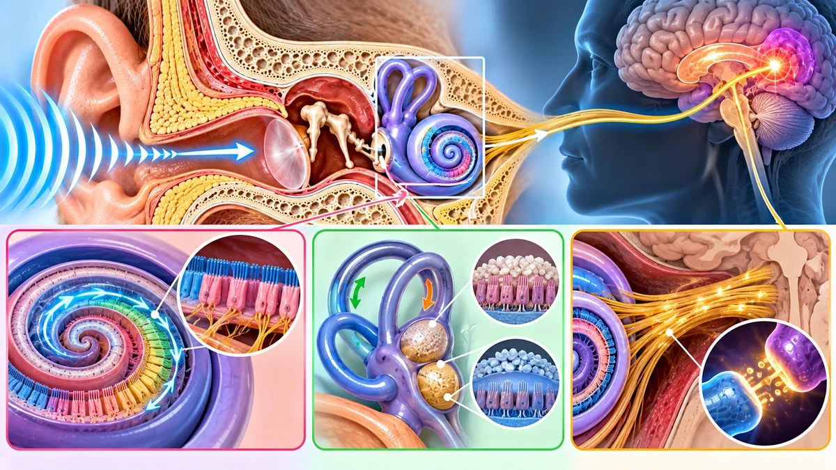
Where does sound actually become something you hear?
The cochlea and its remarkable hair cells
The cochlea is a fluid-filled, snail-shaped structure coiled about two and a half turns inside the temporal bone, and it is where the real transformation happens. A travelling wave moves along the basilar membrane: high-frequency sounds activate hair cells near the cochlear base while low-frequency sounds activate those near the apex, creating a precise frequency-to-location map across the membrane. The hair cells of the organ of Corti convert that fluid movement into electrical signals. When these hair cells are damaged by prolonged noise exposure, ageing or certain medications, the result is sensorineural hearing loss, which is permanent because hair cells do not regenerate.
The vestibular system: balance is an inner-ear job too
Sitting alongside the cochlea are three semicircular canals positioned at roughly right angles to each other, plus the utricle and saccule housed in the vestibule. These structures detect rotational head movement and linear acceleration respectively, sending balance information to the brain at exactly the same time as the hearing signal. Conditions like Menière's disease and vestibular neuritis affect this system, causing vertigo and imbalance even when the cochlea itself is not the site of the problem.
The auditory nerve's final relay
The auditory nerve (part of the vestibulocochlear nerve, cranial nerve VIII) carries encoded electrical signals from the cochlea toward the auditory cortex, where your brain finally interprets them as speech, music or noise. Damage along this nerve or in the cortex itself is classed as auditory neuropathy, a less common but clinically significant type of hearing difficulty. When this pathway is disrupted, speech can sound present but scrambled, even when the cochlea is working perfectly.
Which common ear problem affects which part?
Ear region | Condition | Key symptoms |
|---|---|---|
Outer ear | Cerumen impaction (earwax blockage) | Gradual, low-grade muffling; no pain or discharge |
Outer ear | Pain, itching, swelling, sometimes discharge; worse when touching the ear | |
Middle ear | Otitis media | Ear pain, pressure, muffled hearing, sometimes fever; eardrum may perforate |
Middle ear | Otosclerosis | Gradually worsening conductive hearing loss; no pain or discharge; tinnitus may occur |
Inner ear | Sensorineural hearing loss | Permanent difficulty with speech clarity; not simply a volume problem |
Inner ear | Menière's disease | Episodic spinning vertigo, fluctuating hearing loss, tinnitus, fullness in one ear |
Inner ear (balance nerve) | Vestibular neuritis | Sudden severe vertigo and imbalance; hearing is generally preserved |
Outer and middle ear problems commonly cause pain and muffled hearing; once the underlying cause is treated, hearing usually returns fully because the cochlea's hair cells were never involved. Inner ear conditions follow a very different path. Sensorineural hearing loss is permanent because damaged hair cells cannot regenerate, while Menière's disease produces distinct episodes of spinning vertigo with fluctuating hearing rather than a steady decline. Vestibular neuritis is worth knowing about separately: it produces sudden, severe vertigo and hearing is generally preserved, the key clinical feature that helps distinguish it from Menière's disease.
How do hearing aids work with your ear anatomy?
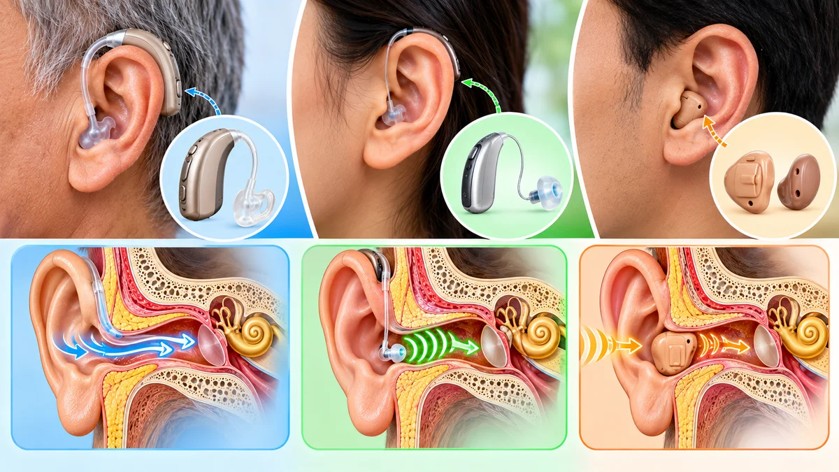
BTE, RIC and ITE styles: which part of the ear does each use?
The style of hearing aid you wear is directly tied to the parts of your ear. Behind-the-ear (BTE) devices sit above the pinna and deliver amplified sound through a tube and earmould that fits inside the ear canal. Receiver-in-canal (RIC) models place the receiver (the speaker) directly in the canal using a thin wire and a soft dome, taking advantage of the canal's natural acoustic properties for a more open, natural sound. In-the-ear (ITE and CIC) devices nestle inside the ear canal itself, making them nearly invisible but requiring a very precise fit to the canal's unique shape, which varies considerably from person to person.
Domes, wax guards and receivers: why the right fit matters
The ear canal produces cerumen constantly, which means every component placed inside it needs regular maintenance. Domes, the soft silicone tips that hold the receiver in position, typically need replacing every one to three months, according to Amplifon, because they can become stiff, discoloured or misshapen with extended use. Wax guards prevent debris from blocking the receiver and need replacing approximately every two to eight weeks, depending on how much wax your ears produce. When these small parts wear out, sound quality drops noticeably, yet some users do not realise that a simple part replacement is often the fix.
VR Speech and Hearing Clinic sells hearing aid accessories such as domes, receivers and wax guards online. If you are unsure which parts fit your device, our audiologists are happy to advise during a free home hearing test or a home fitting visit, so there is no guesswork involved.
Your guide to the parts of the ear and better hearing
Understanding the parts of your ear makes it far easier to make sense of symptoms, conditions and treatment options. The outer ear captures and funnels sound, the middle ear amplifies it mechanically, and the inner ear converts vibrations into nerve signals your brain can read. Each region has its own set of conditions, its own symptoms and its own treatment path, which is why a proper assessment matters so much more than self-diagnosing from a sudden muffling or an occasional ringing sound.
Watch for these symptoms and get them checked without delay: sudden hearing loss in one ear, severe spinning vertigo, pain behind the ear, or hearing that does not return after a cold has fully cleared. These signs can point to conditions that respond far better to early treatment than delayed intervention. Conditions like Menière's disease and sudden sensorineural loss, for instance, are much better managed when assessed within days rather than weeks.
If any of this has made you wonder whether your own hearing is quite right, book a free home hearing test with us at VR Speech and Hearing Clinic. Our certified audiologists visit you at home, assess your hearing thoroughly and can let you trial a hearing aid for a full seven days before you commit to anything. Getting a clear picture of what is happening across each part of the ear is the most useful first step you can take.
Frequently Asked Questions
How do your free home hearing tests work and what do they include?
We carry out free home hearing tests with our certified audiologists visiting you at home to assess your hearing thoroughly and to advise on fittings. We can also let you trial a hearing aid for a full seven days and advise on accessories such as domes, receivers and wax guards during the visit.
Can earwax build-up cause permanent hearing loss?
No, earwax build-up itself does not cause permanent hearing loss and is one of the most common and easily fixed causes of muffled hearing. Permanent sensorineural hearing loss arises when cochlear hair cells are damaged by prolonged noise exposure, ageing or certain medications, because those hair cells do not regenerate.
If I have vertigo, how can I tell whether it is an inner ear problem or something else?
Sudden severe vertigo with preserved hearing is characteristic of vestibular neuritis, while Menière's disease causes episodic spinning vertigo with fluctuating hearing loss, tinnitus and a feeling of fullness in one ear. Middle ear problems more commonly cause pain and muffled hearing that usually returns once the underlying cause is treated.
Which hearing aid style uses which part of the ear and why does fit matter?
BTE devices sit behind the pinna and send sound through a tube and earmould into the ear canal; RIC models place the receiver in the canal using a thin wire and soft dome; ITE and CIC devices sit inside the ear canal itself. The canal's shape affects fit and sound, domes and wax guards need regular replacement, and a precise fit is essential for sound quality and comfort.